PERICARDITIS ECG CHANGES - HEART AND TECHY
PERICARDITIS ECG CHANGES-HEART AND TECHY
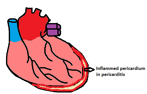
Pericarditis is the inflammation of pericardium.
 |
| Pericarditis |
Acute pericarditis is caused by infective, autoimmune,neoplastic, radiation injury or metabolic causes.
Viral infection is the most common cause of pericarditis.Pericarditis in acute myocardial infarction is due to local pericardial inflammation.Main ecg changes in pericardial effusion are PR segment depression and ST segment elevation
ST segment elevation is occur due to change in epicardium,this exerts a pressure in our heart which will result in decreasing contractility,decrease filling and decrease cardiac output.Our heart consist of 3 layers, epicardium myocardium and endocardium,epicardium is the layer which is very close to pericardium,so changes which occur in epicardium in pericarditis will also affect pericardium
Majority of perfusion occur in our heart during diastole.Changes in epicardium during pericarditis will leads to progressive ischemia,so the damage tissue releases intracellular components,mainly the potassium moves inwards that is from epicardia to inner layers so the current is travelling away this will result in negative depolarization so the baseline of our ecg entire heart as a result due to the increased atrial pressure movement of electrolyte occurs in the opposite direction and p wave down slopping occur in ecg. Key points to diagnose pericarditis in ecg are classic presentation of pericarditis is saddle shaped ST segment,ST segment axis will be shifted to +30 to +60 degree.Diastolic injury current leads to TP segment depression which manifest in ecg as j point elevation .Due to repolarization abnormality in early phase of pericarditis T wave becomes taller and peaked.
Spodick sign is an important diagnostic ecg sign.Changes in qrs complex rarely occur and if present it occurs due to underlying cardiac disease.QT interval shortens due to the shortened duration of activation in injured zone
PR depression and ST elevation are seen simultaneously in leads I,II,III a v f and v 2 to v 6.Ecg changes may not be significant in every patient and variations in ecg are common
SOCIAL MEDIA:
YouTube Channel:
https://www.youtube.com/channel/UCRrYRylZdS67_kbATQrLQqA
Instagram:


Comments
Post a Comment